Vertigo Part 1: Why Does the Room Spin? BPPV, Inner Ear Crystals, and Cervicogenic Dizziness
D
Dr. Annunciato Angelini
Lead Chiropractor (23+ Years Clinical Practice)
|June 14, 2024 10 min read
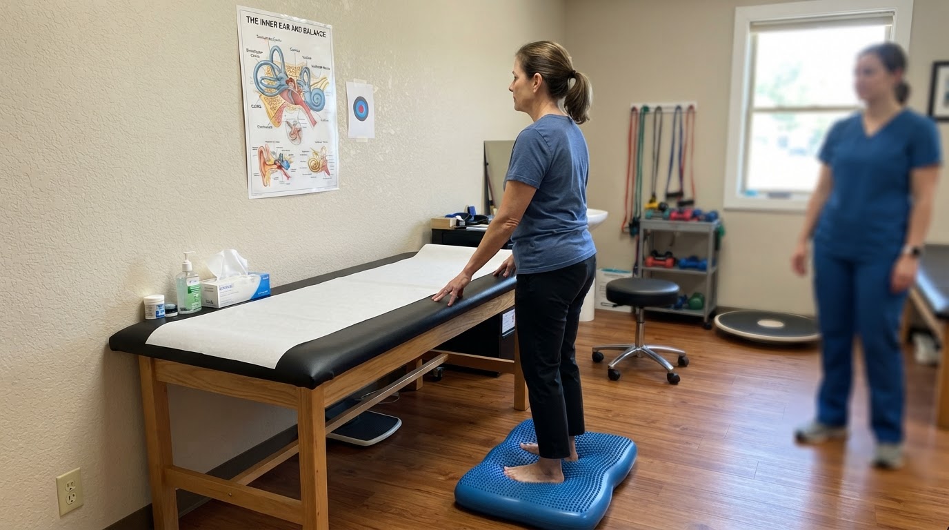
vertigo and BPPV inner ear balance rehabilitation assessment room in Windsor
You wake up in the morning, roll over on your side to turn off your alarm clock, and suddenly the entire bedroom erupts into a violent, sickening spin. The walls whirl around you, your stomach heaves with intense nausea, and you clutch the bedsheets in sheer terror, completely unable to determine which way is up. Even after the violent spinning subsides 30 seconds later, you are left feeling unsteady, 'drunk,' brain-fogged, and terrified to move your head. Vertigo and dizziness are among the most distressing, disorienting, and debilitating complaints treated in clinical healthcare. In this first part of our comprehensive two-part clinical guide on vertigo, Dr. Annunciato Angelini of Active Chiropractic in Windsor explains the physiological mechanisms of equilibrium, the classic presentation of Benign Paroxysmal Positional Vertigo (BPPV), and how neck dysfunction causes Cervicogenic Dizziness.
Understanding Equilibrium: The Three Senses That Keep You Balanced
True balance is not a single sense like sight or hearing; it is a complex, continuous computational masterpiece performed by your central nervous system. Your brain stem and cerebellum maintain equilibrium by constantly cross-referencing sensory data streamed from three distinct anatomical systems:
The Visual System: Your eyes provide a visual horizon and track relative movement in your environment.
The Vestibular System: The delicate inner ear organs that detect gravitational pull, linear acceleration, and angular head rotation in three dimensions.
The Proprioceptive System: Millions of microscopic mechanoreceptors embedded inside your muscles, tendons, and spinal joints—with the highest concentration located in your upper cervical spine (neck)—that tell your brain exactly where your head and limbs are positioned in space.
As long as all three systems send identical, harmonious data to the brain, you feel completely steady and centered. However, if one system begins broadcasting false, conflicting, or erratic data—such as your inner ear screaming that you are somersaulting at high speed while your eyes see a stationary bedroom—the sensory mismatch triggers acute vertigo, violent nystagmus (involuntary eye twitching), nausea, vomiting, and profound balance loss.
What Is Benign Paroxysmal Positional Vertigo (BPPV)? The Loose Crystal Mystery
By far the most common cause of acute, room-spinning vertigo is Benign Paroxysmal Positional Vertigo (BPPV), accounting for over 50% of all vestibular presentations. To understand BPPV, we must explore the vestibular labyrinth inside the temporal bone of your skull. The inner ear contains two otolith organs (the utricle and saccule) and three fluid-filled semicircular canals (anterior, posterior, and horizontal) oriented at 90-degree angles to each other.
Inside the utricle sits a gelatinous membrane studded with thousands of microscopic calcium carbonate crystals called otoconia (commonly called 'inner ear crystals'). These heavy crystals act like tiny paperweights, bending sensory hair cells to tell your brain when you are accelerating forward in an elevator or car. Due to head trauma (such as a concussion or whiplash accident), inner ear viral infections, prolonged bed rest, or natural age-related degeneration of the gelatinous bed, these microscopic calcium crystals can break loose.
Once free-floating, the crystals migrate into one of the fluid-filled semicircular canals—most commonly the posterior semicircular canal (in approximately 85% to 90% of cases). When you tilt your head back, bend over to tie your shoes, or roll over in bed, gravity causes these heavy crystals to tumble through the endolymph fluid like stones in a pipe. This creates abnormal fluid currents that deflect the cupula hair cells, sending a violent, false signal of rapid head spinning that lasts 20 to 60 seconds until the crystals settle at the bottom of the canal.
The Dix-Hallpike Test: How BPPV Is Definitively Diagnosed
BPPV cannot be seen on an MRI, CT scan, or blood test. It is diagnosed exclusively through a clinical positional provocation test called the Dix-Hallpike Maneuver:
During this test, Dr. Angelini positions you on an examination table, turns your head 45 degrees toward the side being tested, and swiftly guides you backward into a lying position with your head extended 20 degrees over the edge of the table. If BPPV is present in that ear, after a brief 2-to-5-second latency delay, the patient experiences a sudden burst of vertigo and displays a distinct, involuntary torsional up-beating nystagmus (the eyeballs rhythmically rotate and jerk toward the affected ear). Observing this precise eye movement confirms the diagnosis and identifies exactly which semicircular canal is contaminated.
Watching a patient go from completely incapacitated by room-spinning vertigo to completely clear and stable after performing a targeted canalith repositioning maneuver is one of the most rewarding moments in clinical practice.
Cervicogenic Dizziness: When the Spinning Stems from Your Neck
While BPPV originates in the inner ear, a huge percentage of patients suffering from chronic, lingering dizziness actually suffer from Cervicogenic Dizziness (cervical vertigo). The upper cervical spine (specifically the C1 atlas, C2 axis, and C3 vertebrae) possesses the highest density of muscle spindles and articular mechanoreceptors in the entire human body.
These cervical mechanoreceptors feed directly into the vestibular nuclei of the brainstem via the cervico-ocular reflex (COR), which coordinates head movement with eye tracking. When the upper cervical vertebrae become subluxated, jammed, or arthritic—or when deep suboccipital muscles lock in chronic spasm from whiplash, poor desk posture, or sports impacts—the mechanoreceptors fire corrupt positional data to the brainstem. The brain perceives a continuous mismatch between neck position and vestibular signals, creating persistent feelings of floating, swimming, rocking on a boat, visual tracking disorientation, and 'drunken' unsteadiness.
If your dizziness is triggered specifically by rolling over in bed and involves violent room-spinning for under 60 seconds, it is likely BPPV. If your dizziness is a constant, vague floating or swimming feeling accompanied by neck stiffness and headaches, it is likely cervicogenic dizziness.
The takeaway
Vertigo is a symptom, not a diagnosis. Whether your spinning is caused by dislodged inner ear crystals in BPPV or corrupt sensory data from upper cervical joint subluxations, identifying the exact anatomical origin is the key to complete, permanent recovery.
Related Care & Clinic Guides
Explore how our team supports patients recovering from these specific conditions in Windsor:

 Soft Tissue & Sports